Brian P. Helmke, Ph.D.

About
Brian Helmke researches the relationship between cell mechanics and cell function using new tools in materials science and molecular biology, with a focus on cardiovascular disease.
Living cells and tissues adapt to their environment by altering structure, gene and protein expression, and biochemical functions. For example, endothelial cells lining the artery wall at the blood tissue interface experience fluid mechanical forces that vary with time and location along the artery. However, the mechanisms by which cells transduce mechanical stimuli into biochemical signals are not well understood. Our laboratory employs a multidisciplinary biomedical engineering approach to understand the relationship between intracellular mechanics and cell function.
Education
B.S.E., Bioengineering, University of Pennsylvania, 1992
B.S.Econ., The Wharton School, University of Pennsylvania, 1992
Ph.D., Bioengineering, University of California, San Diego, 1996
"Our lab employs a multidisciplinary biomedical engineering approach to understand the relationship between intracellular mechanics and cell function."
Selected Publications
Courses Taught
Awards
Featured Grants & Projects
-

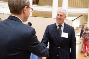
Faculty Spotlight: The Award-Winning Professor With Dad Jokes, Candy Demos and an Umpiring Legacy
Read NowDistinguished professor is just one of the many sides of biomedical engineering’s Brian Helmke.
-
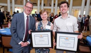
The Class of the Classroom: Biomedical Engineering Professor Wins Accolades for Teaching and Service
Read NowBrian P. Helmke won the 2024 UVA Alumni Association's Distinguished Professor Award.
-

Biomedical, Mechanical and Aerospace Engineering Faculty Members Win Annual Teaching Award
Read NowBrian Helmke and Natasha Smith were recognized as outstanding and inspiring educators with The Jefferson Scholars Foundation’s Hartfield Excellence in Teaching Award.
-
UVA Engineering Shines With Top University Awards
Read NowThree faculty, Brian Helmke, Natasha Smith and Garrick Louis, and graduate student Zackary Landsman were named among UVA’s best for teaching and public service.
-
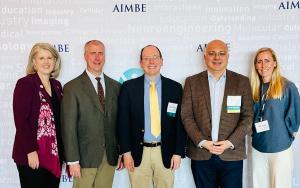
Three UVA Biomedical Engineering Professors Inducted to Acclaimed Medical and Biological Engineering Organization
Read NowBrian Helmke, Timothy Allen and Mete Civelek join 20 other BME faculty as AIMBE Fellows.