The ejection fraction is a standard measure of heart function. It’s the percentage of blood leaving the heart each time it beats. But in terms of diagnosing heart disease, it’s what economists might call a lagging indicator.
“The problem is that ejection fraction is retrospective,” said Frederick Epstein, professor and chair in the Department of Biomedical Engineering at the University of Virginia. “By the time function declines enough to alter the ejection fraction, the damage may be done.”
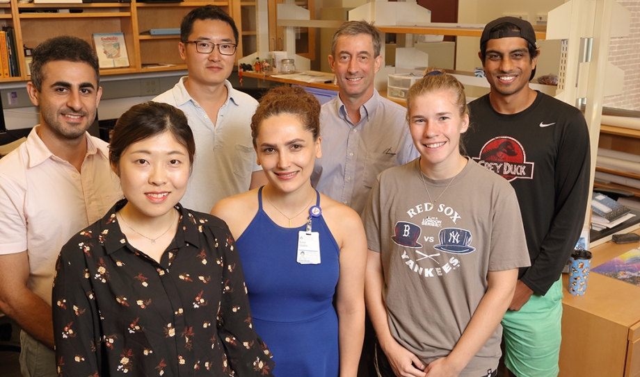
For the last 15 years, Epstein and the researchers in his group have been the world leaders in developing a cardiac magnetic resonance imaging technology that can directly measure the contractile function of heart muscle tissue. The technique is called cine displacement encoding with stimulated echoes (DENSE) strain MRI.
Cine DENSE strain imaging could make it possible for cardiologists to detect early heart dysfunction and intervene early, before damage that could otherwise lead to heart failure. The National Heart, Lung, and Blood Institute at the National Institutes of Health recently awarded Epstein a grant that will move the technology significantly closer to commercialization.
“In many cases, researchers have demonstrated that cine DENSE MRI is a more sensitive indicator than ejection fraction,” Epstein said. “Our plan is that in the future, when patients come in to the clinic for an MRI, cine DENSE cardiac strain imaging will be among the suite of options available to them.”
“We are getting really close.”
The new grant will allow Epstein to tackle the last major set of issues that stand in the way of widespread clinical application. Currently, patients must hold their breath while the image is being acquired, which can be difficult for some patients. Epstein and his team are using the grant to develop a process that separates respiratory motion from cardiac motion, creating accurate images while allowing patients to breathe freely. They are also developing deep learning methods to accelerate and automate the image analysis process.
Finally, they will test their improved technology against the older version in a clinical area the lab has been pursuing, the use of cine DENSE MRI to help cardiac electrophysiologists make better decisions about the use of pacemakers and implantable cardioverter defibrillators.
“Our goal has been to develop an efficient, easy-to-use, accurate and reproducible technique that will be broadly useful for cardiac imaging and other clinical purposes,” Epstein said. “After 15 years, we are getting really close.”

The Backstory
When Epstein first began working with cine-DENSE MRI for cardiac strain imaging, a research team at the National Institutes of Health had created a proof in principle for the technology, but it was limited to a single image of a fully contracted heart. “Heart imaging is not very useful unless you can do time-resolved imaging over the cardiac cycle,” Epstein said. “They stopped at a single image because they knew how difficult getting beyond that point was going to be.”
Cine DENSE MRI is indeed a complex technology rooted in the physics of how tissue displacement modifies MRI signals. Simply put, as the heart contracts and tissues move, it causes a phase shift in the MR signal that can be imaged and the displacement calculated. “We get extremely accurate measurements of motion, and from that calculate the strain—the deformation of the heart during contraction,” Epstein said.
Epstein and his lab have moved the technology forward on a number of fronts. They found a way to create a sequence of time-resolved images over the cardiac cycle and developed computational methods to calculate strain maps from the MR images. They also invented a number of methods to reduce artifacts in the images and improve the signal-to-noise ratio.
Today, companies like Siemens and Circle CVI, which specializes in cardiac imaging software, are already working to bring the technology to the imaging suite. Meanwhile, research institutions from around the world—including Stanford, UCLA, Emory, the Royal Brompton Hospital in London and the Université de Lyon in France—are exploring such potentially groundbreaking applications for cine DENSE MRI as monitoring cancer patients for chemotherapy-induced cardiotoxicity, detecting cardiac remodeling and dysfunction caused by childhood obesity and spotting cardiac dysfunction in diabetes patients.
Not all of the potential applications of cine DENSE MRI involve the heart. One research group at Johns Hopkins is considering cine DENSE MRI for planning brain surgery, while another is using these methods to assess the health of cartilage in the knee.
“We make our research readily available and encourage others to use it,” Epstein said. “By working with all these groups around the world, we have been able to amplify our impact ten-fold.”