What if doctors could predict a breast cancer patient’s response to treatment in just two weeks? Researchers at the University of Virginia have turned that possibility into reality, with a pioneering method that uses “zero-passage” organoids — tiny, patient-specific tumor models — to fast-track testing of therapies.
Luminal breast cancer, the most common subtype, is defined by its reliance on hormones like estrogen to fuel growth. While these cancers are often less aggressive, they pose a challenge in the lab — conventional cell cultures often fail to retain the unique traits of the patient’s tumor.
By speeding up the process of figuring out how a tumor reacts to treatment, this new method gives doctors a powerful tool to personalize care by cutting down on trial-and-error, minimizing side effects, and giving patients a better shot at beating their cancer.
How It’s Done
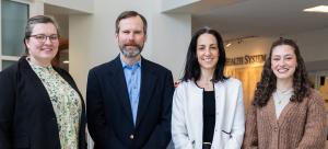
Team members Róża Przanowska, a biomedical engineering research associate; Najwa Labban, a biomedical engineering M.D.-Ph.D. student in the Medical Sciences Training Program; Kevin Janes, the John Marshall Money Professor of Biomedical Engineering; and Shayna Showalter, professor of surgery, collaborated with a multidisciplinary team of surgeons and pathologists to integrate their research seamlessly into the clinical workflow.
The team used scrapes of tumors collected immediately after a patient undergoes surgery to create 3D organoids — miniature replicas of the tumor cells that remain viable and responsive to different treatments in the laboratory yielding results in 14 days.
Their work was recently published in Breast Cancer Research.
Showalter explained, “We are working at the intersection of surgery, pathology and engineering to create a novel system that could dramatically change how we treat cancer. This method will eventually allow doctors to offer individualized treatments to patients based on their specific tumor.
Pathologists collaborated to standardize tissue handling and provide expert input on tumor viability. Biomedical engineers then optimized organoid culture techniques, allowing dozens of replicate organoids to be tested with different therapies simultaneously.
The team achieved success rates twice as high as previous organoid models, enabling them to test drug and genetic therapies on real patient tumors with speed and precision. “This collaboration allowed us to bridge the gap between the operating room and the lab,” Przanowska said. “We’ve created a workflow that retains the individual characteristics of the tumor cells while being scalable for basic and translational research.”
Why It Matters
Early-stage luminal breast cancers require years of follow-up to determine if treatments are working. This method offers a new way to shorten that timeline by providing oncologists with fast, personalized insights into a tumor’s likely response to therapy.
“Our approach eliminates months of guesswork,” Labban said. “This is about delivering actionable data directly to the clinical team.”
By focusing on patient-derived material, this method could help oncologists predict which therapies will work best for individual patients. It also minimizes exposure to ineffective treatments, reducing side effects and improving quality of life.
“This kind of work doesn’t happen in isolation,” Janes said. “It’s a testament to what can be achieved when clinicians and scientists come together with a shared goal. By combining our expertise, we’ve created something that could have a profound impact on cancer care.”
Publication and Grant Information
The study, titled “Patient-Derived Response Estimates from Zero-Passage Organoids of Luminal Breast Cancer” and published in Breast Cancer Research on Dec. 31, 2024, features contributions from researchers at the University of Virginia across several departments.
The core research team authors included Róża K. Przanowska, Ph.D., research associate; Najwa Labban, M.D.-Ph.D. candidate; and Kevin A. Janes, Ph.D., John Marshall Money Professor of Biomedical Engineering, all of the Department of Biomedical Engineering at the UVA School of Engineering and Applied Science; and Shayna L. Showalter, M.D., professor of surgery in the UVA School of Medicine.
Additional authors were Piotr Przanowski, research scientist, and Russell B. Hawes, M.D.-Ph.D. student, both of the Department of Biomedical Engineering at the UVA School of Engineering and Applied Science; and Kristen A. Atkins, M.D., professor of pathology in the UVA School of Medicine. Acknowledged members of the clinical team at the UVA School of Medicine were David Brenin, Lynn Dengel and Anneke Schroen in the Department of Surgery; and pathology staff members Kristan Abernathy, Biljana Amanovic, Sarah Bastarache, William Lee Carstens, Lisa Friedman, Tappy Gish, Akriti Gupta, AnnaVi Jones, Michelle Kline, Lauren Luketic, Shannon May, Nicole McGinn, Margaret Moore, Lauren Pelkey, Ansley Scott and Selveras Zayed.
This work was supported by several grants, including NIH research grants (U54-CA274499 and R01-CA256199); pilot and training awards from the UVA Comprehensive Cancer Center’s Breast Translational Research Team (GR014313) and the UVA Farrow Fellowship (PJ03500); and NIH training-transition awards (K00-CA253732, T32-CA009109, T32-GM145443 and T32-GM007267).
Additional support was provided by the UVA Center for Systems Analysis of Stress-adapted Cancer Organelles (SASCO); resources from the UVA Flow Cytometry Core Facility and Research Histology Core Facility, partially supported by the UVA Comprehensive Cancer Center Support Grant (P30-CA044579); computational resources from UVA Research Computing; and Emily Farber and Suna Onengut-Gumuscu from the UVA Center for Public Health Genomics.