Jeff Saucerman, Ph.D.

About
Our lab combines computational modeling and high-throughput experiments to discover molecular networks and drugs that control cardiac remodeling. Our experimental approaches include high-throughput microscopy and -omic profiling of various types of primary and induced pluripotent stem cell (iPSC)-derived cardiac cells (e.g. cardiomyocytes, fibroblasts, macrophages). Our computational approaches include large-scale modeling of signaling/gene regulatory networks, machine learning on -omic data, and mining of electronic health records. Specific application areas include:
- cardiac hypertrophy, survival, and regeneration
- cardiac development
- inflammation-fibrosis coupling
Education
Ph.D. in Bioengineering, University of California San Diego, 2005
B.S. in Engineering Science, Pennsylvania State University, 2000
We are mapping the complex networks that control heart function and failure.
Research Interests
Selected Publications
Courses Taught
Awards
Featured Grants & Projects
-
UVA Engineering Researchers Develop AI Tool To Fast-Track New Health Treatments
Read NowUsing a new AI tool, biomedical engineering professor Jeff Saucerman’s team discovered a drug used by millions for anxiety and depression also has surprising heart-healing potential.
-
Biomedical Engineering Professor Receives $3.1M NIH Grant to Improve Heart Attack Outcomes
Read NowJeff Saucerman will collaborate with researchers at the School of Medicine to study the role of lipoprotein receptors in heart failure.
-
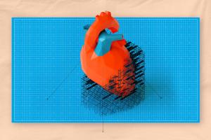
How Do You Mend a Damaged Heart? UVA Researchers Have Solid Leads
Read NowBiomedical engineering professor Jeff Saucerman and his research team have identified medications that might be able to regenerate heart cells to repair damage.