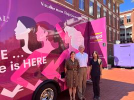
Earlier this month, Dr. Kelsey Kubelick, faculty in the Department of Biomedical Engineering at the University of Virginia, welcomed Seno Medical Instruments to Grounds for a hands-on demonstration of their groundbreaking Imagio® Breast Imaging System. As part of Seno’s national “Roadshow,” the team travels the country in a vibrant pink RV, bringing their FDA-approved imaging technology directly to academic and clinical communities.
Dr. Kubelick organized and hosted the event, in collaboration with Dr. Rachelle Crescenzi (Associate Professor in the Department of Radiology and Medical Imaging at the University of Virginia), which drew faculty, clinicians, and students from across UVA to both the Main Hospital and Fontaine Research Park. Attendees explored the capabilities of the Imagio® system through interactive stations, including operating the device on tissue-mimicking models. The demonstration showcased how optoacoustic/ultrasound (OA/US) imaging could revolutionize breast cancer diagnostics and inspire new research directions in biomedical imaging.
A New Frontier in Breast Cancer Imaging
Breast cancer imaging remains a complex challenge in clinical diagnostics. Traditional methods like mammography and ultrasound often yield high false-positive rates, leading to unnecessary biopsies and patient anxiety. The Imagio® system offers a noninvasive alternative that combines optoacoustic imaging with ultrasound to deliver molecular and cellular-level insights—far beyond what conventional anatomical imaging can provide.
Optoacoustic imaging (also known as photoacoustic imaging) uses laser pulses to stimulate tissue, generating sound pressure waves that are captured to form detailed images. Unlike mammography, OA imaging avoids harmful X-rays and patient discomfort, while offering high optical contrast that can detect cancer at its earliest clinically significant stage.
Seeing Beneath the Surface
What sets OA/US apart is its ability to visualize blood-based signals, enabling analysis of vascular growth, density, and morphology in and around tumors. This deeper insight into tumor biology, microcirculation, and metabolic state is being leveraged to:
- Identify molecular subtypes of breast cancer
- Guide personalized treatment planning
- Evaluate response to therapy
Looking Ahead
The demonstration highlighted the potential of OA/US imaging not only to enhance breast cancer diagnostics but also to open new research pathways in biomedical imaging and beyond. Dr. Crescenzi played a key role in supporting and leading the Day 2 session at Fontaine, further emphasizing the collaborative effort across departments to bring cutting-edge imaging technology to UVA. As technologies like Imagio® continue to evolve, they promise to reshape how clinicians detect, diagnose, and treat breast cancer—offering hope for earlier detection and better outcomes.