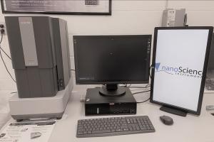
Thermo Scientific Phenom XLG2 Scanning Electron Microscope (SEM)
About
Location: Jesser Hall room 139
The SEM microscope is capable of imaging and microanalysis on all specimens, with or without preparation. Characterization of fractures and polished sections from metals, semiconductors, and ceramics. Attachments for elemental analysis with Energy-Dispersive X-ray Spectroscopy (EDS).
SEM Technique Summary:
- SEM and Compositional Analysis of Metals, Glasses, Semiconductors, Ceramics, Polymers, Geologic Materials, Catalysts, Pharmaceuticals, etc.
- High vacuum imaging: <0.25 mTorr
- Low-vacuum imaging: 0.25 mTorr to 1.5 Torr
- Environmental SEM (ESEM): 1.5 Torr to 6 Torr
- Quantitative Elemental Analysis and Mapping of Materials with EDS
- EDS with Oxford 50 mm Silicon drift detector (SDD) sensitive down to boron
- EDS Elemental Sensitivity:> 0.1 wt.% (weight percentage) for Z > 5
- Max magnification: 200,000x
- Sample size up to 4 x 10 x 10 cm³ ( H x W x D)
Phenom XLG2 Features:
- CeB6 source for electrons
- Accelerating voltage: 5, 10, 15, & 20 kV
- <10 nm imaging resolution at 30 kV and 10 nm resolution at 3 kV
- Secondary electron imaging (SEI) for topographic contrast
- Backscattered electron imaging (BEI) for chemical contrast
- High vacuum, low vacuum and Environmental SEM (ESEM) capability
- 100 mm stage travel in both the x and y directions
- Three available stages: standard (36-sample), large sample (100 mm x 100 mm x 40 mm), and eucentric tilt stage (6-axis)
Instrument Training Videos from Nanoscience can be found on YouTube. These include:
- How to Load a Sample on a Phenom Desktop SEM | Episode 1
- How to Load a Sample on a Phenom Desktop SEM | Episode 2
- How to Navigate Samples on a Phenom Desktop SEM | Episode 3
- How to Optimize SEM Images on a Phenom Desktop SEM | Episode 4
- How to Optimize SEM Images on a Phenom Desktop SEM | Episode 5
- Exploring Electron Beam Settings on a Phenom Desktop SEM | Episode 6
- Detector Options on a Phenom Desktop SEM | Episode 7
- How to Enhance EDS on a Phenom Desktop SEM | Episode 8
- ParticleMetric Analysis on Phenom Desktop SEM | Episode 9
Additional details about the Phenom, including an introductory applications and sales video, can be found on Youtube.
Specific information about the Phenom XLG2 and 3D Roughness Reconstruction software, can be found on the Thermo-Scientific website.
Rates
UVA RATES:
Phenom XLG2 SEM Instrument Usage: $45/hr
Phenom XLG2 Operator Costs: $60/hr
EDUCATIONAL, GOVERNMENT & NON-PROFIT INSTITUTIONS RATES:
Phenom XLG2 SEM Instrument Usage: $55/hr
Phenom XLG2 Operator Costs: $70/hr
INDUSTRIAL PARTNER RATES:
Phenom XLG2 SEM Instrument Usage: $200/hr
Phenom XLG2 Operator Costs: $125/hr