Optical Microscopy
The NMCF maintains a number of optical (light) microscopes to magnify features on samples, including a Hirox digital microscope for enhanced depth-of-field images and video. Our Zygo white-light profilometer provides quantitative measurements of surface roughness and topography. Raman spectroscopy utilizes UV and visible laser light to identify molecules and study chemical bonding between atoms and molecules on specimen surfaces, generating spectra for single points or surface maps with micron resolution. An integrated Atomic Force Microscope (AFM) allows supplemental determination of topography and electrical characteristics.
NMCF Optical and Raman Instrumentation
-
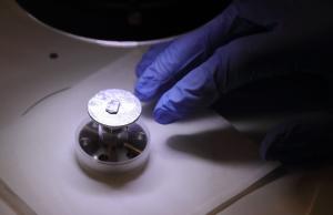
Renishaw Raman Spectrometer with Integrated AFM
Confocal Raman microscope and integrated AFM provides nondestructive chemical composition and phase information at micron-level resolution, in conjunction with topographic, nano-mechanical and electronic properties. Hot/Cold and heating stages available for temperature-programed spectroscopy.
-
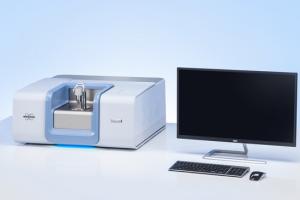
Bruker FTIR Spectrometer
The NMCF Bruker Fourier-transform infrared spectrometry (FTIR) provides nondestructive compositional information of materials (solids, powders, films, and liquids) based on molecular vibration information, both molecular identification and quantification. Attenuated total reflectance (ATR) and diffuse reflectance assessories are available.
-
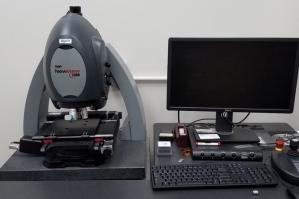
Zygo White-light Profilometer
White-light optical profilometry is utilized for determination of surface topography and measurement of surface shape, surface finish, surface profile roughness (Ra), or in surface area roughness (Sa), surface texture, asperity and structural characterization.
-
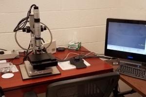
Hirox Digital Microscope
The NMCF Hirox Microscope provides nondestructive digital imaging, videography, and measurement of material surfaces of all kinds. Stage tilt for inspection of surface roughness and stage rotation for object shape information and 3-D modeling are available.
-
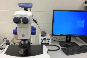
Zeiss Discovery Stereomicroscope
The Zeiss Stereo Discovery V8 Steromicroscope provides optical brightfield, darkfield, and oblique imaging via transmitted or reflected light for visual inspection of materials at 25-200x. Cross polarizers allow identification of isotropic or anisotopic features, and a long focal length allows easy sample manipulation. An 8-megapixel camera captures high-resolution images and video.
Contact Us

Joe Thompson
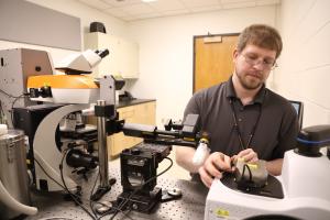
Joe Thompson joined the NMCF as a Laboratory Specialist, also providing analysis for the Virginia Department of Transportation (VDOT). He specializes in Raman Spectroscopy, Atomic Force Microscopy, and Optical and Electron Microscopies.

Catherine Dukes
Catherine Dukes directs the Laboratory for Astrophysics and Surface Physics (LASP) and provides expertise in X-ray photoelectron spectroscopy (XPS) for the University of Virginia's Nanoscale Materials Characterization Facility (NMCF). Her NSF and NASA funded research focuses on the interaction of radiation with surfaces.