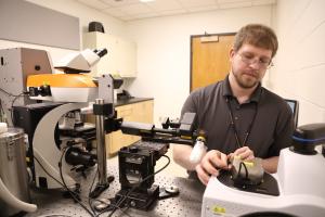
Raman Spectrometer-Atomic Force Microscope (Raman-AFM)
Molecular Identification and Chemistry of Surfaces, combined with Nano-Scale Surface Morphology

About
Location: Jesser Hall room 142
Renishaw InVia™ Confocal Raman microscope and integrated Bruker Innova AFM provides nondestructive chemical composition and phase in conjunction with topographic, nano-mechanical and electronic properties. Raman mapping with 1 micron spatial resolution. Tip-Enhanced Raman spectroscopy (TERS) for enhanced surface specificity. Photoluminescence spectroscopy. Hot/Cold and Heating stages available for temperature-programmed spectroscopy.
Raman Spectroscopy Technique and Specifications Summary:
- Non-destructive Molecular Compositional Analysis of Pharmaceuticals, Biostructures, Semiconductors, Photovoltaics, Polymers, Geology, Mineralogy, Pigments, Contaminant Identification, etc.
- Molecular Composition of Solids, Liquids, and Loose or Pressed Powders
- Identification of Molecular Impurities with High Sensitivity
- Raman does not provide information on Metals and Alloys
- Molecular Information Depth: ~ 1micron, ~10 nm with TERS
- Spatial Resolution: < 2 micron, ~ 20 - 30 nm with TERS
- Spectral Range: 405 nm - NIR (limit is 1022 nm or 1.213 eV)
- Laser Line Cut-Off: Approx 100 cm-1 from the laser line (except for 405 nm laser which has a usable cutoff approx. 200 cm-1 from the laser line)
- Collect sequential Raman and AFM measurements
- Hot Stage Temps: Room Temp to 1773K (1500 C)
- Hot/Cold Stage Temps: ~120K - 873K ( -196 - 600 C)
- Sample size: from <10 micron dia. to 5’ x 3’ x 8” (size of optical table)
Atomic Force Microcopy Technique and Specifications Summary:
- Non-destructive analysis of Sample Surface Topography
- Modes: Contact mode, Tapping Mode and STM Available
- Samples maybe in air or in liquid
- Single Point Spectroscopy for a detailed characterization of local electrical or mechanical properties
- Magnetic Force Microscopy (MFM) measures magnetic force gradient distribution above the sample surface
- Electric Force Microscopy (EFM) measures electric force gradient distribution above the sample surface
- Nanolithography with probe tip to scribe or indent a sample surface by mechanical pressure
- AFM Mapping space: 250 x 250 x 100 microns3
- Sample size: to 45mm x 45mm x18mm
Raman Features:
- Full system automation (laser, grating, and filter selection)
- 4 Wavelengths available from 3 Lasers:
- Diode Laser: 785 nm
- Argon Laser: 514 nm
- Argon Laser: 488 nm
- Diode Laser: 405 nm
- Microscope with 4 objectives: 5x, 20x, and 50x, plus 50x with large numerical aperture
- Three gratings: 1200gr/mm, 1800gr/mm, and 3000gr/mm
- Stereo viewing (binocular eyepieces)
- Point mapping, StreamHR, depth profile, volume mapping
- Automated measurement queuing
- Automated Raman calibration correction (quick calibration)
- Laser auto-align
- Raman signal auto-align
AFM Features:
- Large Area scan: XY >90 µm, Z >7.5 µm (Closed-Loop)
- Small Area scan: XY >5 µm, Z >1.5 µm (Open-Loop)
- Open-Loop Drift: <1 nm/min
- Closed-Loop Drift: <3 nm/min (warm-up time 15 min)
- Images of height (scan size 1 to 10 microns) and feedback enabled, scan rates are 1-4 Hz
- Images size: 16 x 16, 32x32, 64x64, 128x128, 256x256, 512x512 or 1024x1024
- Motorized Z-travel: 18mm
- On-axis, 1.25 mm - 0.25 mm FOV
- Software-controlled 5x motorized zoom
- 10x objective (50x optional)
- <2 µm resolution (0.75 µm resolution with 50x)
Contact Us

Joe Thompson
Laboratory Specialist, Materials Science & Engineering and VDOT
NMCF Principal Scientist for Raman, AFM, SEM/Optical Microscopy, & Metallurgy
Joe Thompson joined the NMCF as a Laboratory Specialist, also providing analysis for the Virginia Department of Transportation (VDOT). He specializes in Raman Spectroscopy, Atomic Force Microscopy, and Optical and Electron Microscopies.